Volar Plate Injuries by Lucille Roberts
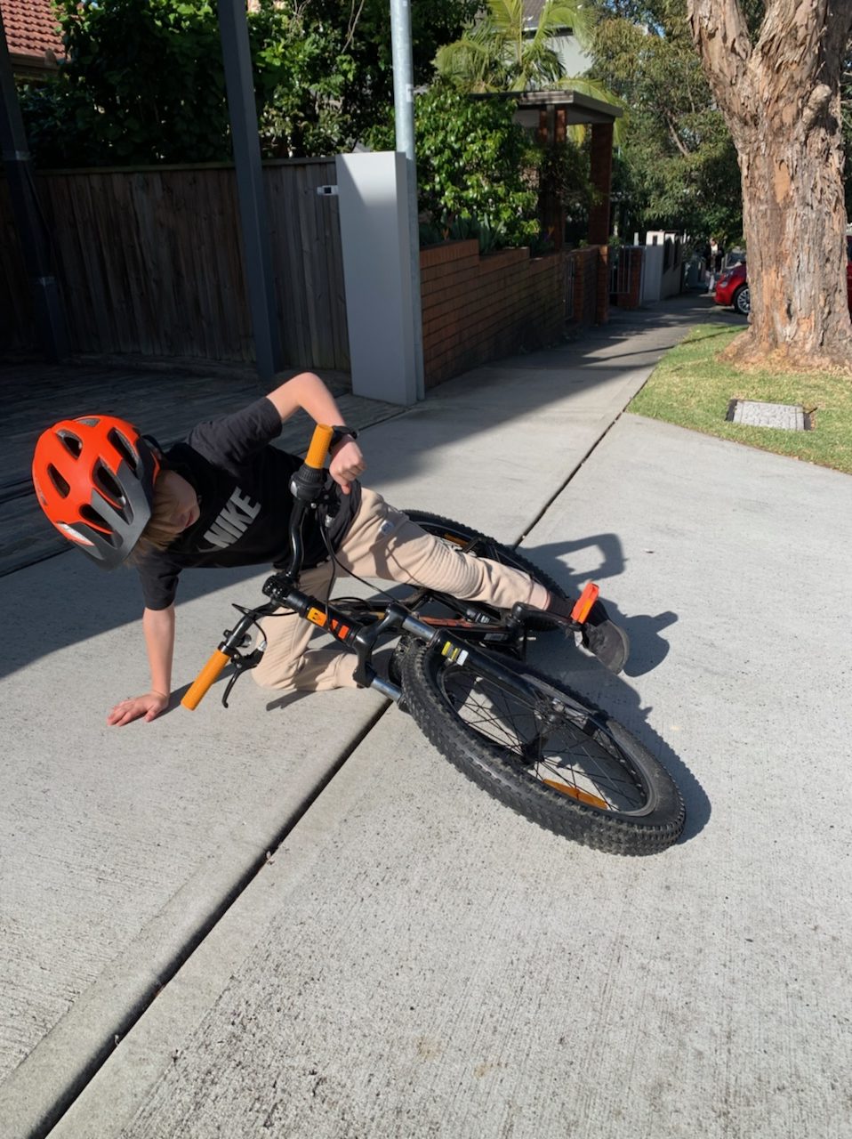
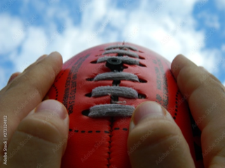
The volar plate is susceptible to damage when the finger receives a hyperextension force, commonly a result of a ball injury (e.g. basketball, netball).
Anatomy: Together with other ligaments it is vital in preventing hyperextension. The Volar Plate forms the floor of the proximal interphalangeal joint (PIPJ), joining the proximal and middle phalanges together on the volar side of the PIPJ.
Injury: When the PIPJ is forced into hyperextension the volar plate can become sprained or detached, it is more common for it to detach distally off of the middle phalanx.
If the volar plate is avulsed proximally, off of the proximal phalanx, it can be interposed in the joint and prevent the joint from being relocated, requiring a surgical consultation.
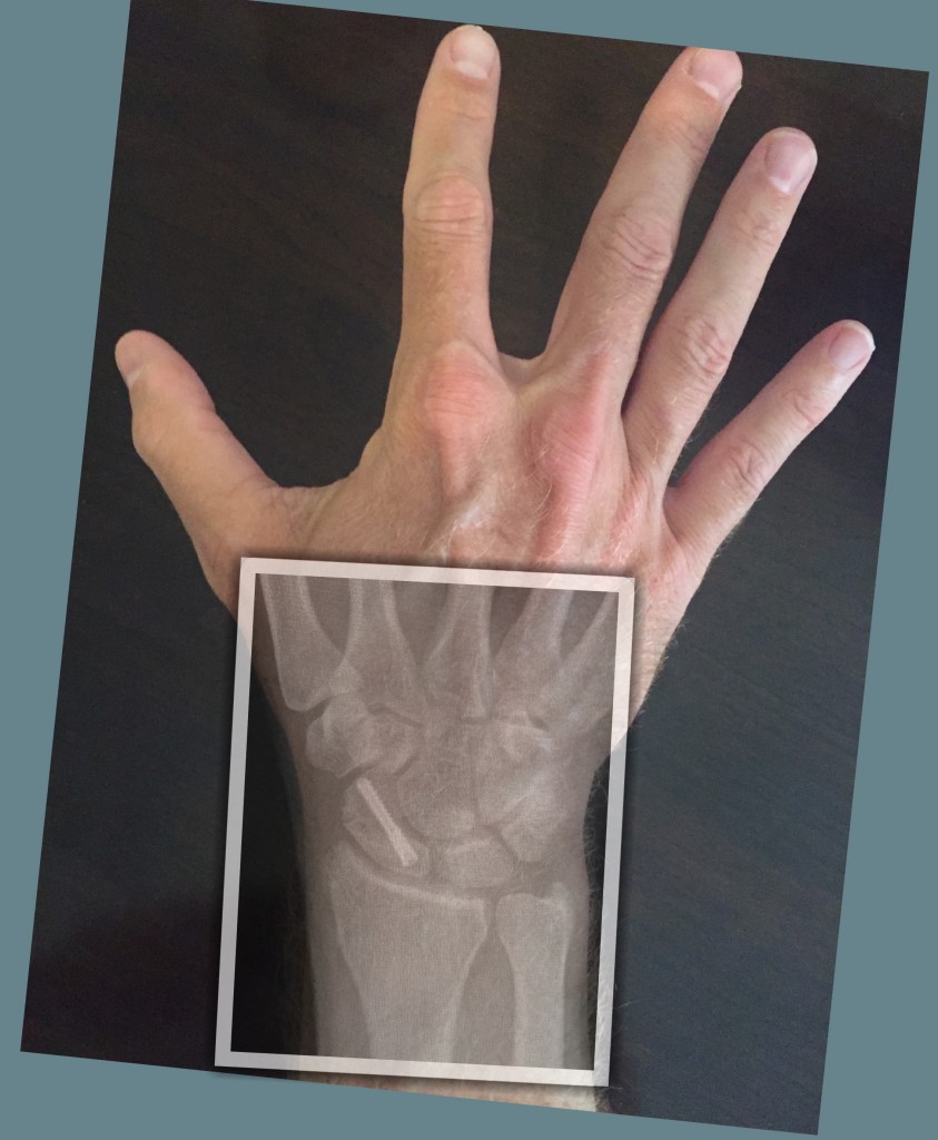
X-ray: Radiographs may be clear if the volar plate is sprained or ruptured. The volar plate can also become detached with or without a bony fragment. If there is a bony avulsion, the size of the fragment is important; if it is greater than 40% of the articular surface the joint may be unstable and require a surgical consultation. Small avulsion fractures are usually successfully treated conservatively.Treatment:
Treatment
Initial Splint: Volar plate injuries can be stabilised within a splint. The proximal interphalangeal joint (PIPJ) needs to be splinted in slight flexion for the Volar Plate to securely re-attach to its distal connection. A dorsal thermoplastic splint is fabricated with the PIPJ in approximately 30 degrees flexion. For sprained volar plates buddy taping may be adequate support.
Exercises: are commenced immediately within comfort in the splint, avoiding extension beyond the limit of the splint. Flexion is usually limited by pain and swelling, however can progress quickly, ideally most patients will have a full fist at the completion of splinting.
Oedema management: is an early priority, and reducing localised swelling enables better movement. Often Coban is applied under the splint, and patients can be taught how to re-apply this as their swelling reduces.
Splinting: the angle of flexion is reduced as the PIPJ feels stable in extension. Splinting is usually ceased between four to six weeks. Dependent on the patients needs they may then move to a neoprene finger stall, buddy taping or no splint at all.
Strengthening: patients can be helped to regain full strength, assisting in confidently returning to functional hand use.
The aim of hand therapy is to provide appropriate management of the injury and return patients to full movement, strength and function as soon possible.
Why do we care so much?
As I am writing this article a patient has attended for an unrelated injury on their other hand, and as often happens during the course of the appointment they have shown me an old injury and asked if anything can be done. Unfortunately it’s too late for this finger. The injury happened approximately 15 years ago and received no treatment.
Photos show patient trying to straighten little finger. The middle joint hyperextends and the end joint flexes – Swan Neck Deformity.
There are a few injuries that can cause a swan neck deformity (SND), untreated Mallet fingers being one of them, volar plates that do not reattach are another. On assessment this patient could actively straighten his tip so I could rule out an untreated mallet finger. The PIPJ could be hyperextended very easily with no firm end feel. The client now finds that each time they straighten their little finger it jumps into the SND position. This is uncomfortable and irritating. Thankfully they can make a full fist and their job is not typing!
This is why we care so much about what can appear to seemingly small injuries; they can have lifelong negative impacts for our patients on a daily basis.
We want our patients to be healthy and active and back on that netball or basketball court as soon as possible, by providing appropriate and timely care for an injured finger hand therapy can do that!